|
|
|
|
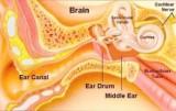
External EarConsists of pinna and the external auditory meatus up to the lateral border of the tympanic membrane.
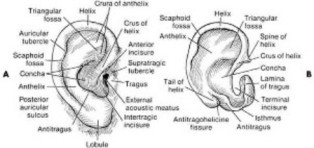
PinnaComposed mostly of cartilage and has no useful muscles. The center, concha, leads to the EAM. Resonance : 5kHx Resonances and antiresonances. Differentiate sound sources in front of the listener from those behide.
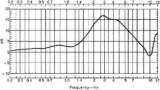
External Auditory MeatusLateral 1/3 : cartilaginous portion containing cerumen-producing glands and hair follicles Remaining 2/3 : bony portion, including a tight dermal lining surrounding the TM Resonance : determined by the length of the tube.
Behaves like a quarter wave resonator. 2.5 cm à 3.5 kHz Reasons for NIHL.
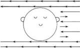
External EarHead and external earLocalization of sound sources Attenuator at frequencies at which the width of the head is greater than the wavelength of the sound : 2Hz
Lower frequencies Interaural time differences : 0.6 msec Higher frequencies Useful for improving the detection and recognition of low-energy, high-frequency sounds such as voiceless fricatives. Hearing-aid and evaluations
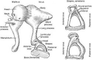
Middle Ear
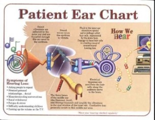
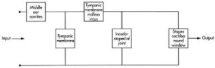
The middle ear consists of the tympanic cavity and the osseous eustachian tube. Transmits acoustic energy from the air-filled EAM to fluid-filled cochlea.
Impedance-matching 3 ways
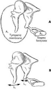
Pressure gain : 25-30 dB Tympanic Membrane
Transformer action of TM and ossicular chain provides for a relatively efficient transfer of power to the inner ear and the fidelity of sound transmission across the middle ear is outstanding. Distortion of signal does not occur even >130 dB SPL Passive mechanical system with both mass and compliant elements and, therefore, resonant properties Linear system coupled to the cochlear which contributes a large resistance. Highly damped and linear and has a wide frequency response Ratio of volume velocity of the stapes to sound pressure at the TM increases in humans to about 800-900 Hz – the resonance of middle ear. (50% loss is only 3 dB) Less than half of the power entering the middle ear reaches the cochlea. Inefficient at frequencies above 2kHz The reason humans do not detect and recognize sounds above 20 kHz The frequency region of greatest energy concentration is 3-5 kHz 2 striated muscles : tensor tympani and stapedium
Protection function of the muscles
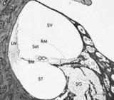
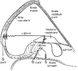
CochleaA coiled, bony tube about 3.5 mm long. Divided into the scala vestibuli, scala media and scala tympani : extracellular fluidlike material Endolymph : intracellular-like fluid Scala media has a positive DC RP of about 80 mV, decreasing slightly from the base to apex : produced by the heavily vascularized stria vascularis of the lateral wall “The Battery of cochlea” (Na+/K+-ATPase pumps).
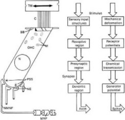
Acoustic energy pathway
Basilar membrane
Organ of Corti
Outer and inner hair cells of the organ of Corti play a major role in the transduction of mechanical energy (acoustic) into electrical energy (neural) The spiral ganglion
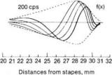
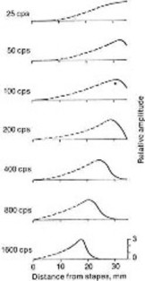
Displacement pattern of the basilar membrane is traveling wave
Cochlear amplifier
Stereocilia
The potassium flux
Neurotransmitter
Gross Cochlea PotentialEndolymphatic (endocochlear) potentialDC, 80-10 mV, Scala media Not generated in response to acoustic stimulation Cochlear microphonic potentialAC, K+ current flow through outer hair cells Summating potentialDC, several origin, reflects the DC shifts Whole nerve action potentialAll-or-non discharge of auditory nerve
Gross Cochlear PhysiologyMalfunctioning of the mechanism involved in the production of endolymph and the EP can produce metabolic presbycusis. When the flow of endolymph is blocked, endolymphatic pressure is increased and hydrops in produced
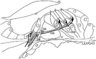
Eighth Nerve Physiology30,000 neurons that innervate the cochlea
90-95% synapse directly on inner hair cell Bipolar, myelinated Each inner hair cell is innervated by about 15-20 type I neurons Type II neurons : outer spiral fibers5-10% innervate the outer hair cells Monopolar, unmyelinated Each type II neuron branches to afferent innervation pattern of the cochlea originating from SOC projection to the cochlea Spontaneous rate
The most sensitive fibers have the most spontaneous activity The most basic measure of auditory nerve function is the tuning curve of a single fiber. The tuning curve
Normal neural activity, including sensitivity and frequency-resolving power, depends on intact outer hair cells and normal stereocilia One of the most common features of SNHL is recruitment of loudness.
Nonlinear Properties of the EarThe outstanding features of the cochlea and the auditory nerve Two-tone rate suppression Otoacoustic emission (OAE)
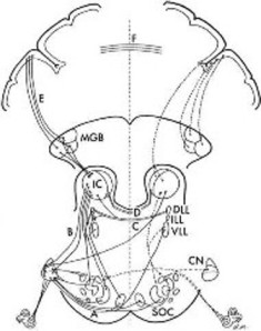
Auditory Central Nervous System
Summarized By Thirayost Nimmanon â´Â ¸ŐĂÂĘ¶ě ąÔÁÁŇąą·ě
|
|
»ĂŃş»Ăا¤ĂŃé§ĹčŇĘŘ´ 15/06/2010
ä´éĂŃşˇŇĂʹѺʹع Web Hosting ¨Ňˇ SPAComputer.com, ThaWang.com |