|
|
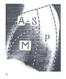
MediastinumCentral cavity of the thorax Sides : pleural cavities (parietal pleura) Inferior : diaphragm Superior : thoracic inlet Anterior : sternum Posterior : vertebral body No compartment of body carries more physiologic traffic.
CompartmentsSuperiorSuperiorly, the fascial planes communicate with the neck à Infection spreading AnteriorAnterior : sternum Posterior : upper dorsal spine Inferior : anterior border of heart MiddleSuperior : anterior border of heart Inferior : diaphragmatic surface Posterior : anterior border of dorsal spine Posterior
ContentsAnterior-Superior : thymus gland, aortic arch, SVC, internal mammary vessels, lymph node, parathyroid gland, ectopic thyroid tissue. Middle : pericardium, heart, great vessels, trachea, tracheal bifurcation, main bronchi, phrenic nerve, hilar lymph node. Posterior : esophagus, vagus nerves, sympathetic chain, thoracic duct, thoracic desending aorta, azygos and hemiazygos vein, paravertebral lymph node.
Tumors and Cysts25% are primary cystic lesions. Thymic neoplasm is the most common primary tumor. Lymphoma, neurogenic, germ cell tumors are common tumor. In childhood series, malignancy could be found 50% of tumors, which are Hodgkinĺs disease or Non-Hodgkin Lymphoma. Lymphoma is the most common malignancy in all age.
Clinical Manifestations1/3 of Patients are asymptomatic. Most common symptoms are nonspecific (chest pain, cough, dyspnea) Compression of adjacent structures : trachea, esophagus, Superior vena cava, recurrent nerve palsy, Hornerĺs syndrome Endocrine syndrome Hypertension : pheochromocytoma Hypercalcemia : parathyroid tumor Thyrotoxicosis : intrathoracic goiter Gynecomastia : choriocarcinoma Systemic consequence : Pel-Ebstein fevers in Hodgkinĺs disease
95% of incidental radiographic findings are benign. In symptomatic patients, 50% are found to be malignancy. In age <2 years old. 87% of benign present with tracheal obstruction. Poorer prognosis is associated with Hornerĺs syndrome, vocal cord paralysis, hemiplegia.
DiagnosisChest X-ray : localizing lesion. CT (+ oral or IV Contrast) : It is most diagnostic of benign pathology. Appearance of solid malignancies is less definitive. Malignant characteristics are readily demonstrated. MRI It has benefits for vascular lesions, Heart and Great vessels Contraindications :
Barium Swallow : for invasion, compression and displacement of esophagus. Arteriography : is the diagnostic standard for pre-operative evaluation of major vascular disorders. Venous Angiography : reveals extent of involvement and nature of collateral channels in SVC obstruction. Myelography : is for posterior mediastinal tumors (replaced by CT+MRI). Radioisotope : is to visualize the anterior mediastinal mass. Endoscope : is for esophagus and tracheobronchial tree with biopsy Percutaneous transbronchial or transesophageal needle biopsy Mediastinoscopy and parasternal Mediastinotomy : for lymphoma or thymoma Definitive Resection :
Neurogenic Tumor
It arises from sympathetic ganglion or intercostal nerves. It is most commonly found in posterior mediastinum. Incidence : Adulthood, 10-20% are malignant. Higher proportion of childhood are malignant. Pain, caused by nerve compression or bony erosion, is the most common symptom. Hemiparesthesis, hemiparesis, cord compression can be found : ôDumbbellö extension is characteristic in some cases. Hormonally active tumors cause Hypertension, flushing, diarrhea, diaphoresis, anorexia, fever.
Types of neurogenic Tumors
Neurilemoma (Schwannomas)Schwannoma is the most common tumor neurogenic tumor(40-60%). It arises from Schwann cells in intercostal nerves. It is hard, yellowish, well-encapsulated. Most are benign Dumbbell extension could be found but not common.
NeurofibromaNeurofibroma arises from nerve sheath and nerve cells It is 10% of neurogenic tumor. It is Poorly encapsulated, resemble neurilemomas radiographically. If it is found with Von Recklinghausenĺs Disease, risk of malignant degeneration is increased. It has poor prognosis.
NeuroblastomaIt is most poorly differentiated neurogenic tumor. It arises from sympathetic nervous system 10% occur as primary lesion. Vanillylmandelic acid, an active metabolite, causes Hypertension, fever, vomiting, diarrhea. Because it is radiosensitive, debluking followed by RTx is the treatment of choice. It has good prognosis if found in mediastinum 1st year of life.
Ganglioneuroma, GanglioneuroblastomaGanglioneuromaIt arises from mature cells in sympathetic ganglia. It is found in younger age group. Radiographic : triangular configuration. GanglioneuroblastomaIt arises from mixture of mature and immature cells. It found in the age <3 years old.
Paraganglionic TumorsPheochromocytomasIt arises from chromaffin paraganglionic tumors. It can produce catecholamines. It is 1% of all pheochromocytomas. It is more ôsilentö, more often malignant. ChemodectomasIt is non-Chromaffin in origin. It arises from chemoreceptors :
Treatment : Operation :Operation is performed in most of posterior mediastinal neurogenic tumors Incision : Posterolateral thoracotomy CT à intervertebral foramina and vertebral bodies MRI à intraspinal extension Intraforaminal extension à combined thoracic and neurosurgical procedure MalignantExcised if possible NeuroblastomaRadical operations are approached selectively Active PheochromocytomaPre-operative medical management of paroxysmal Hypertension.
Thymoma
Thymoma is the most common tumor in anterior mediastinal mass. It is found second most common among tumors and cysts. Symptoms
There is no characteristic radiographic feature. Diagnosis is provided by excised mass. Types :
Pleural metastases could be found. Surgery : median sternotomy à 65% curative
Lymphoma
50% of Hodgkinĺs disease and non-Hodgkin lymphoma are found in mediastinum. It is the most common mediastinal malignancy. It is found in anterior mediastinum. Treatment : chemotherapy and radiotherapy Resection is never indicated. Surgery is used only for diagnosis or determining residual active tumor after treatment.
Teratodermoid Tumors
It is found in anterior mediastinum. It is composed of multiple tissue type. It arises from bronchial cleft and pouch cells and is associated with thymus. Mediastinum is the second most common location (It is most commonly arise in Gonad) Symptoms
Radiographiclarge, well ľcircumscribed anterior mediastinal mass 20-50% are calcified. CT : fat density in the center of cystic mass AFP and B-hCG à malignancy TreatmentExcision through median sternotomy is best for Diagnosis and treatment. Imcomplete resection is occasionally necessary.
Germ-Cell Tumors
Germ cell tumors is a rare tumor found in anterior mediastinum. Histogenesis is poorly understood. It may arises from pluripotential primordial germ cells. Teratoma is end point of benign differentiation Types
Incidence : Young adult, male>female 4x It is highly malignant. 80-90% are symptomatic. Symptoms :Nonspecific, mass effect Investigations :Posteroanterior and lateral CXR : 90% detected CT Serum tumor markers hCG : chorioCA, seminoma, ECC AFP : ECC, yolk sac Metastasis from gonadal tumor must be excluded by ruling out the retroperitoneal involvement. Treatment
Mesenchymal Tumors
LipomaIt is most common mesenchymal tumor. It has no fixation. It is asymptomatic despite of enormous size.
FibromaMore dense and less common than lipoma.
Tumors of Vascular origin
Uniform density on CXR and cystic mass on CT.
Endocrine Tumors
ThyroidIt spreads by direct substernal extension. Treatment : Radionuclide
ParathyroidIt is approachable through a cervical incision. It is associated with upper pole of thymus.
Mediastinal Cysts
Mediastinal cyst is the most common found in middle mediastinal compartment. Chest X-ray : opaque densities in typical location CT : near water density in characteristic location
Pericardial CystsPericardial cyst is the most common mesenchymal cyst. It is always asymptomatic. It is most commonly found at right cardiophrenic angle, occasionally communicate with pericardium. Treatment : close observation
Bronchogenic CystsIt is located just posterior to carina or main stem bronchi. Air-fluid level found in tracheobronchial tree distinguish bronchogenic cyst from pericardial cyst. Viscid fluid is found in the cyst. Contrast esophagogram is helpful for diagnosis. It is lined by ciliated respiratory epithelium. Surgery : posterolateral thoracotomy
Enteric CystIt is found in posterior mediastinum adjacent to esophagus. Itĺs rarely found to be communicated with esophagus. Itĺs lined by intestinal mucosa, containing clear colorless mucoid fluid. Peptic ulcer is found if itĺs lined by aberrant gastric mucosa. 60% in Patients age <1 years old have symptoms of tracheal and esophageal compression. Chest X-ray, esophagogram, CT with contrast are helpful. Surgery : posterolateral thoracotomy
Summarized By Thirayost Nimmanon ╩├ě╗Ô┤┬ ŞŇ├┬╩Âý ╣ď┴┴Ď╣╣Ěý
|
|
╗├Đ║╗├장├ĐÚž┼ŔĎ╩ě┤ 15/06/2010
ń┤Ú├Đ║íĎ├╩╣Đ║╩╣ě╣ Web Hosting ĘĎí SPAComputer.com, ThaWang.com |